Chapter 1 : The Cells of the CNS

Brain: Contents Page

The Neural Tube and Neural Tube Defects

The Neural Tube

In the earliest stages of development the embryo consists of a ball of cells, within which three layers can be easily identified.

  1. The outer layer, Ectoderm, will develop into the skin and central nervous system.
  2. The innermost layer, Endoderm, will form the lining of the gastro-intestinal tract.
  3. Between these is the Mesoderm, which will form the muscles, bones and many of the organs of the body.

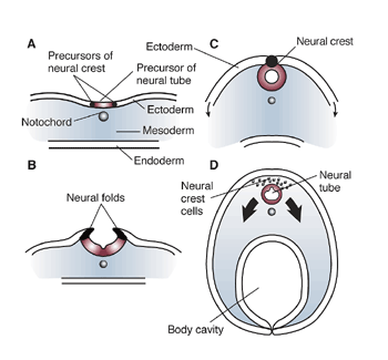
The earliest sign of the nervous system is the development of the neural plate on the dorsal surface of the ectoderm, behind a column of cells known as the Notochord. The notochord is present in all vertebrates and is essential for inducing the development of the neural plate.

At the lateral edges of the neural plate, ridges appear that grow and fold towards each other to form a tube, the neural tube. The cells that lead this development are called Neural crest cells (C), and when they have completed their role in forming the neural tube, they go on to form some more specialised parts of the nervous system including the dorsal root ganglia, the autonomic nervous system and the adrenal medulla (D).

Thus the Neural tube is formed from the primitive Ectoderm of the embryo, and its formation depends on the presence of the notochord; the notochord is also important in the development of primitive nerve cells in the ventral half of the neural tube into motoneurones. The neural crest is important in ensuring the closure of the neural tube, and this process can go wrong leading the Neural Tube Defects.


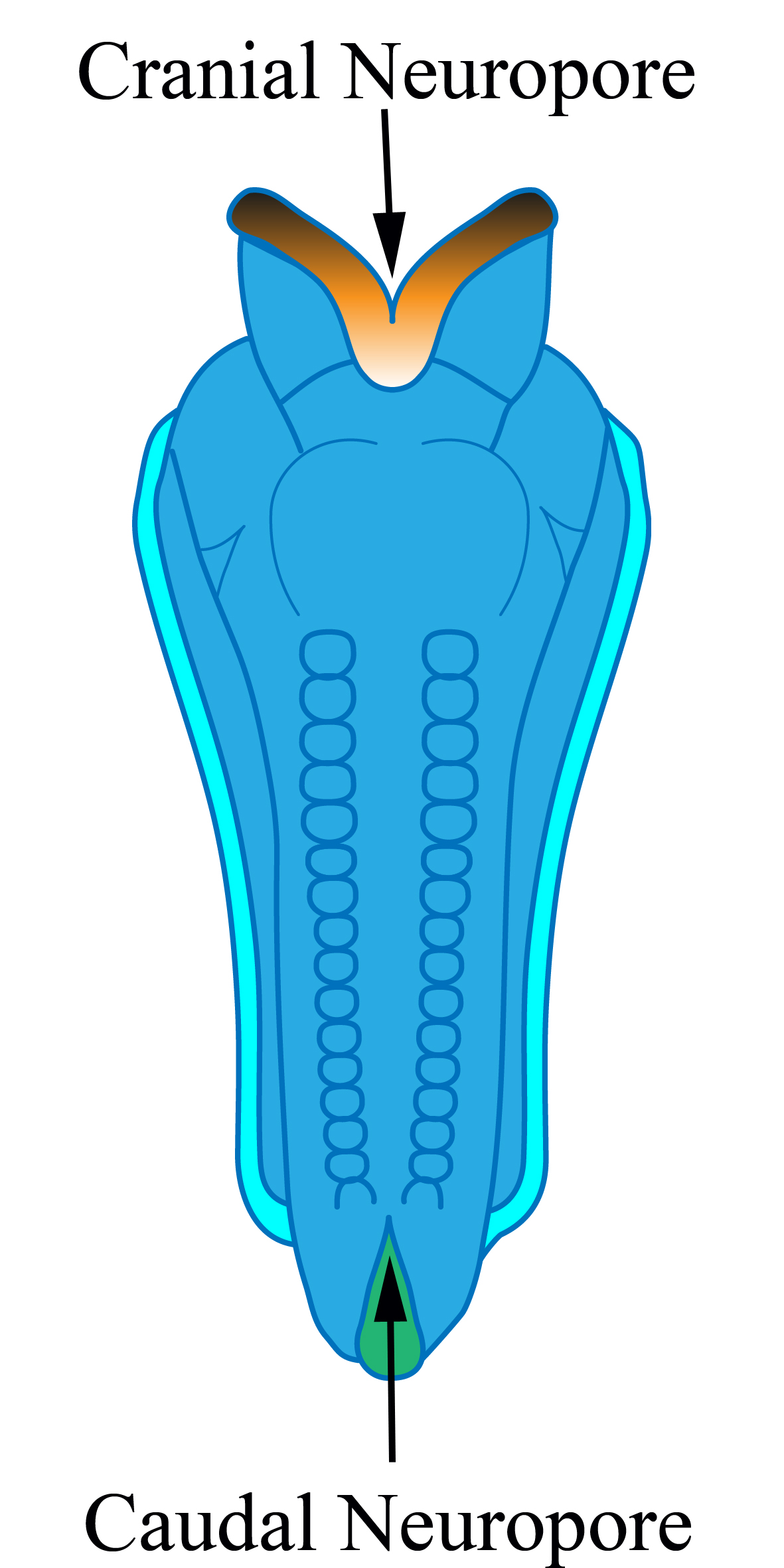
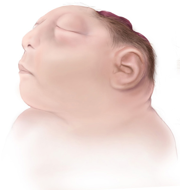
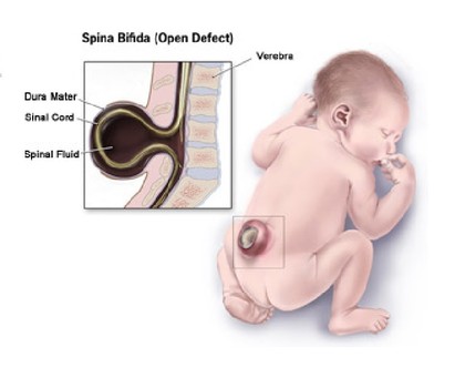
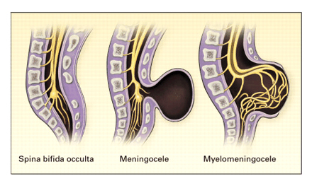
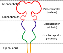
As the neural tube is forming, the central region closes first, and for a short time, the neural tube is open both at both ends, and the openings are called neuropores, these close completely around 28 days in the human.

The diagram shows the cranial and caudal neuropores before closure of the neural tube. Once closed, the neural tube becomes segmented, and each segment is called a somite.

After closure, the cranial end of the neural tube develops into primitive brain which can be divided into four distinct regions:

  • the prosencephalon, which becomes the forebrain
  • the mesencephalon, which becomes the midbrain
  • the rhombencephalon, which becomes the hindbrain and cerebellum, in addition to
  • the spinal cord.

    Anencephaly occurs when the cranial neuropore does not form a complete tube, and the rostral developments of the nervous system fail to occur - so the bably is born without a brain.

    Spina bifida occurs when the caudal neuropore fails to close completely.

  •  

    The cranial end of the neural tube develops into massive structures in the adult human:

    • the prosencephalon, which expands enormously to become the cerebral hemispheres, basal ganglia, thalamus and hypothalamus
    • the brainstem, consisting of the mesencephalon (midbrain) and the rhombencephalon, which becomes the hindbrain to which the cerebellum is attached.

    The spinal cord is segmented with 8 cervical, 12 thoracic, 5 lumbar and 5 sacral segments in humans. When the caudal neuropore fails to close, the deficiency is usually inthing the lumbo-sacral segments of the spinal cord.


    Top


    Neural Tube Defects


    Neural tube defects can be:

    1. Open: The failure of the neural tube to close results in the exposure of neural tissue on the surface of the skin, and is associated with leakage of cerebrospinal fluid on to the skin
    2. Closed: neural tissue is not exposed and is covered by skin, although the skin at this site may not be normal
    3. The commonest neural tube defects develop (A) in the brain:

      • Anencephaly (failure of brain development)
      • Encephalocele (intracranial tissue herniates out through a defect in the skull).
      and (B) in the spinal cord:
      • Spina bifida
      • Myelocoele
      • Meningomyelocoele

      These differ in the extend of the neurological lesions.

      Anencephaly is not compatible with life.

    *

    Anencephaly

    Spina Bifida

    In spina bidifda occulta, there are defects in the vertebral column and maybe some discolouration of the overlying skin, but the nerves remain in roughly their normal position.


    In a meningocoele there is a herniation of the meninges through a gap in the back of the lower segments of the vertebral column. The nervous elements however do not enter this space.


    In meningomyelocoele, the spinal roots and sometimes the cord herniates into this space. In the diagram, 1 represents the CSF within the meningomyelocoele, and 2 indicates the nerves entering the meningomyelocoele..

    If the skin overlying this lesion breaks down , CSF leaks out and infection can easily enter the spinal canal, with disastrous consequences.


    Incidence of Neural Tube Defects

    Neural tube defects now occur in about 0.8/1000 total births.

    Anencephaly and spina bifida account for up to 95% of all neural tube defects.

    Predisposing factors:

    Family history of malformation, chromosomal disorders and deficiency of folic acid (or treatment of the mother with some anti-epileptic drugs that are also folic acid antagonists) all predispose to neural tube defects.

    *

    Meningomyelocoele

    *
    Types of Spina Bifida

    http://som.flinders.edu.au/FUSAWikis/dsrswiki01/doku.php?id=spina_bifida_biological_aspects
    Uvainio via Commons.Wikipedia
    http://www.cdc.gov/ncbddd/birthdefects/anencephaly.html

    Top


    Brain: Contents Page

    © HumanPhysiology.Academy 2014-2015